A Journey into the Microscopic World: Observations of River Water and Pollen Germination
Microscopic study of river water and pollen germination
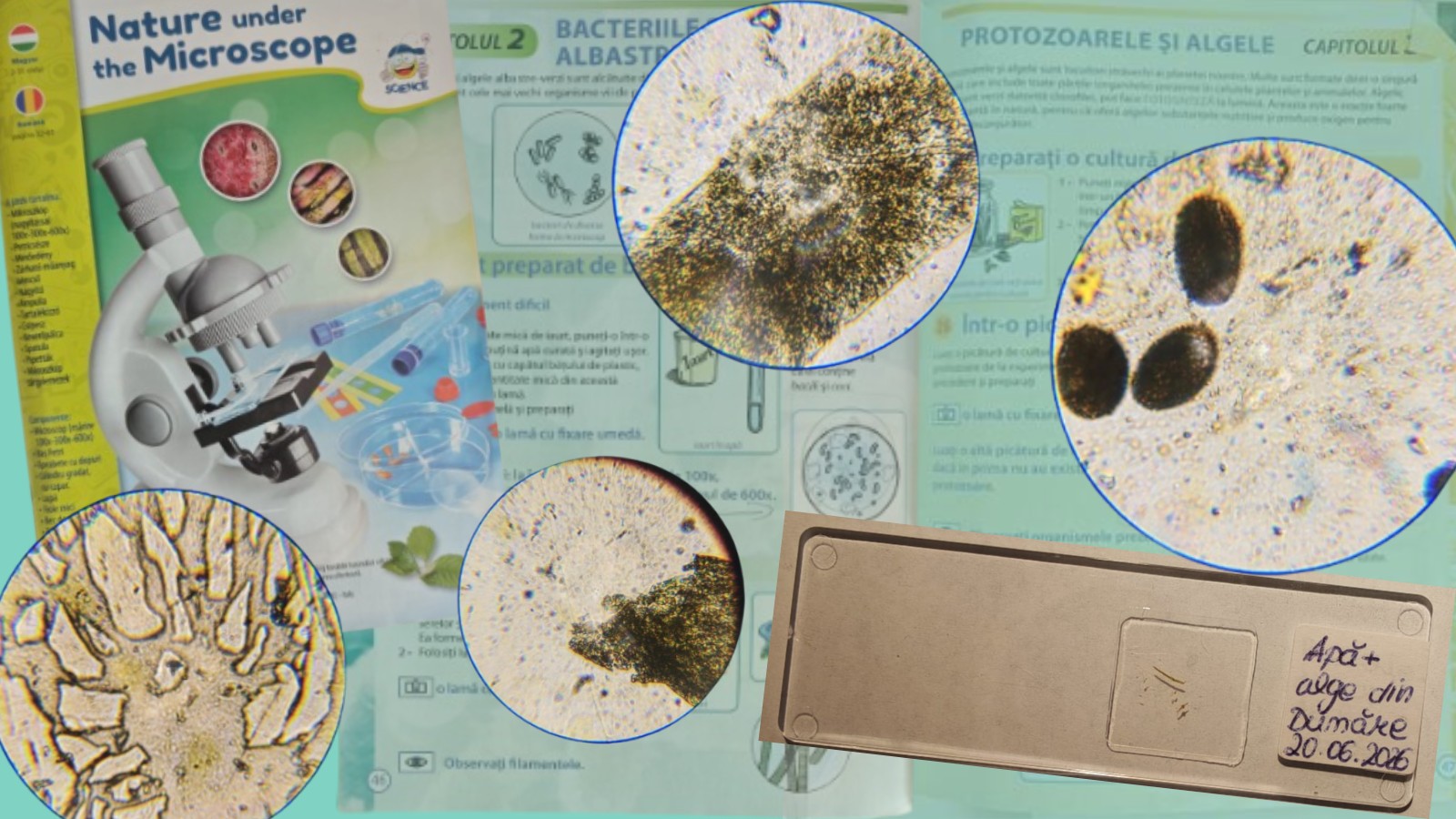
This week, I continued exploring one of my hobbies: microscopy. Interested in the natural world, I kept experimenting with observations under the microscope using a guide I have been following. In particular, I wanted to revisit Experiment 38, which focuses on pollen germination, as I was curious whether I could obtain different or clearer results compared to my previous attempt.
While going through the guide, I also came across an experiment I had always wanted to try: observing green algae under the microscope and possibly studying them in a simple home setup without laboratory equipment. For this purpose, I collected a water sample during a boat trip on the Danube, which also contained visible traces of algae floating in the water.
I am aware that the Danube area includes protected ecosystems, especially for species such as yellow and white water lilies, which are endangered. For this reason, I made sure to collect only water from open areas, avoiding any disturbance of aquatic plants or surrounding habitats.
After one week, I examined the sample. Upon opening the test tube containing the river water and algae, I noticed a very strong, pungent and unpleasant odor, likely caused by natural decomposition processes. To safely handle the sample, I used gloves and two protective face masks during the experiment.
Microscopic observation of river water and algae
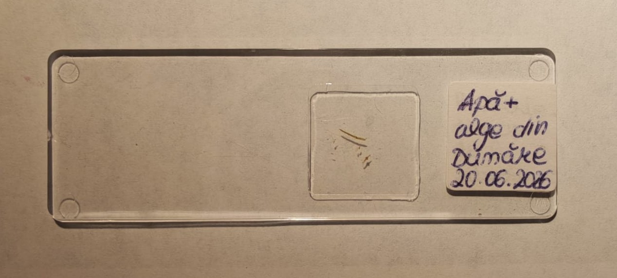
First, I placed a drop of river water onto a microscope slide using the wet mount technique. Simply put, a wet mount is a microscopy method for observing liquid samples using a cover slip to improve clarity under the microscope.
I also isolated a small portion of the algae sample, as it appeared to have small particles attached to its surface, possibly sediment or microorganisms from the river environment.
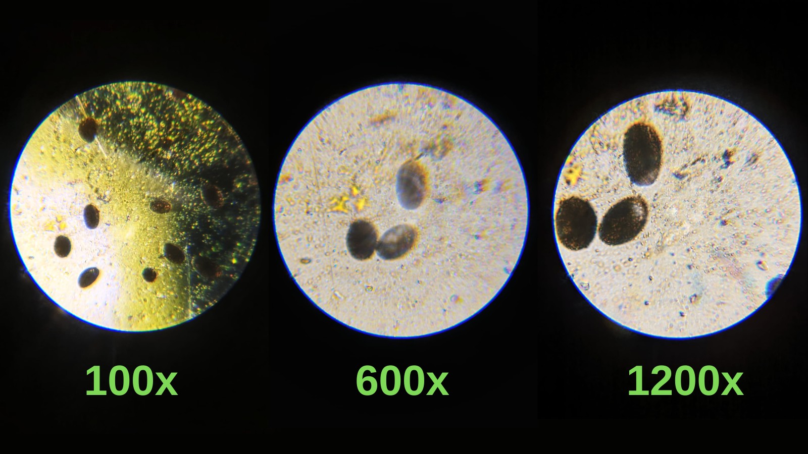
I took photos at each magnification level (100×, 600×, and 1200×) using my phone.
At lower magnification, a green filamentous structure was visible, which I identified as the algae sample. Around it, there were small particles and sediment from the riverbed, which is expected in natural water samples. At higher magnification, a transparent structure became more visible, containing small yellowish dots arranged in a pattern that resembled a chain or ladder-like structure. While I cannot precisely identify its nature, it appeared to be part of the algae’s internal or attached structure and was particularly interesting to observe.
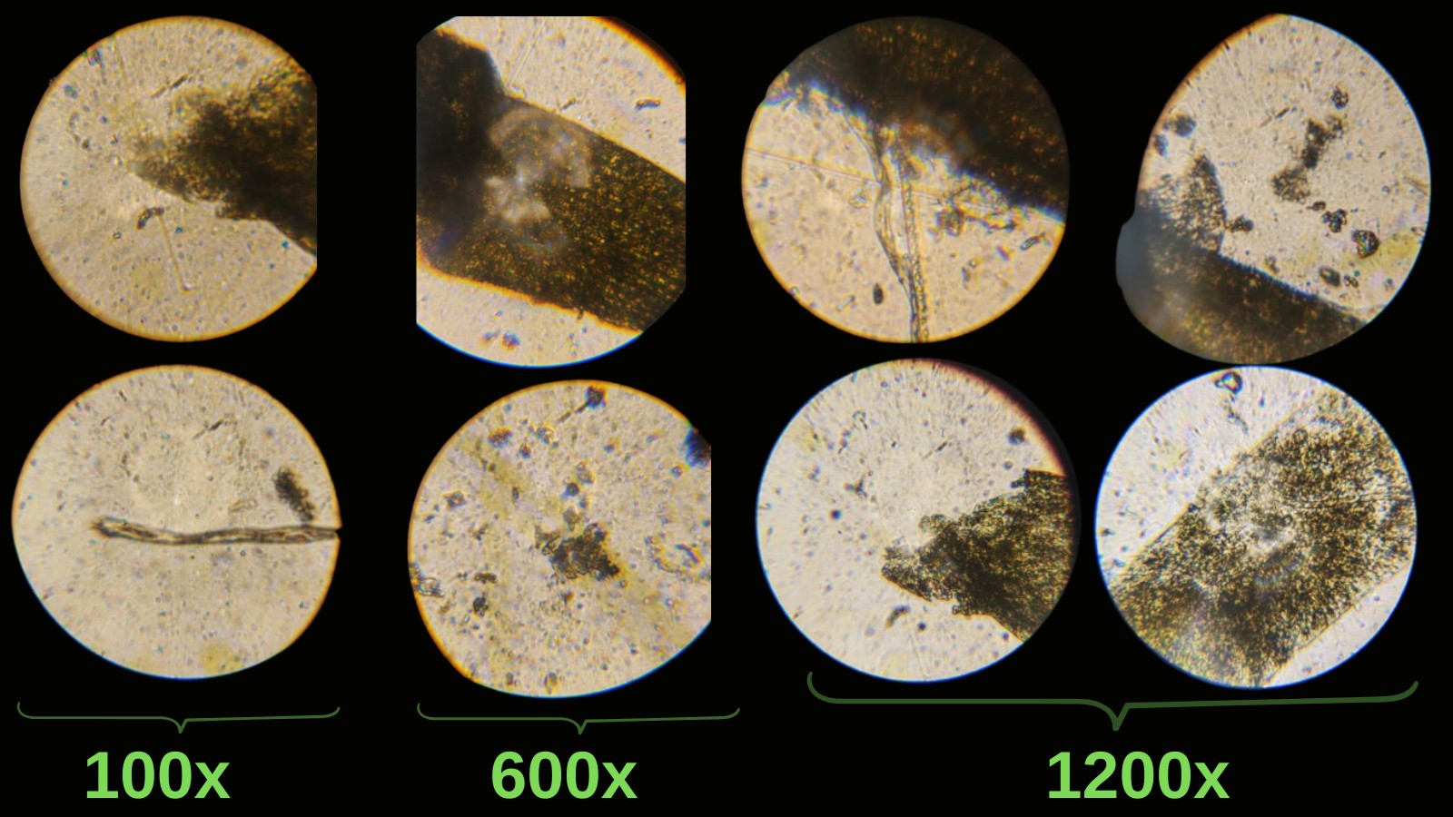
I had also hoped to observe more diverse or “ancient” forms of algae, similar to those found in more isolated environments, but the sample mainly reflected a typical river ecosystem. Nevertheless, it provided valuable microscopic detail.
Pollen germination experiment
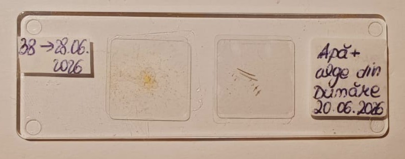
The second part of the experiment focused on pollen germination. Following the instructions in the guide, I prepared a sugar solution by dissolving sugar in water. I then collected pollen from lilies, plants known for their strong fragrance and highly visible pollen grains, and mixed it into the solution. The sample was left to sit for approximately three hours.
After this period, I placed a drop of the solution onto a wet mount slide. The solution had taken on an orange tint due to the pollen pigments.
Under the microscope, I observed multiple brown oval structures, which I identified as pollen grains. At higher magnification, smaller yellow particles were visible around some of the pollen grains. Based on their appearance, these may correspond to pollen germination structures, although this remains a qualitative observation.
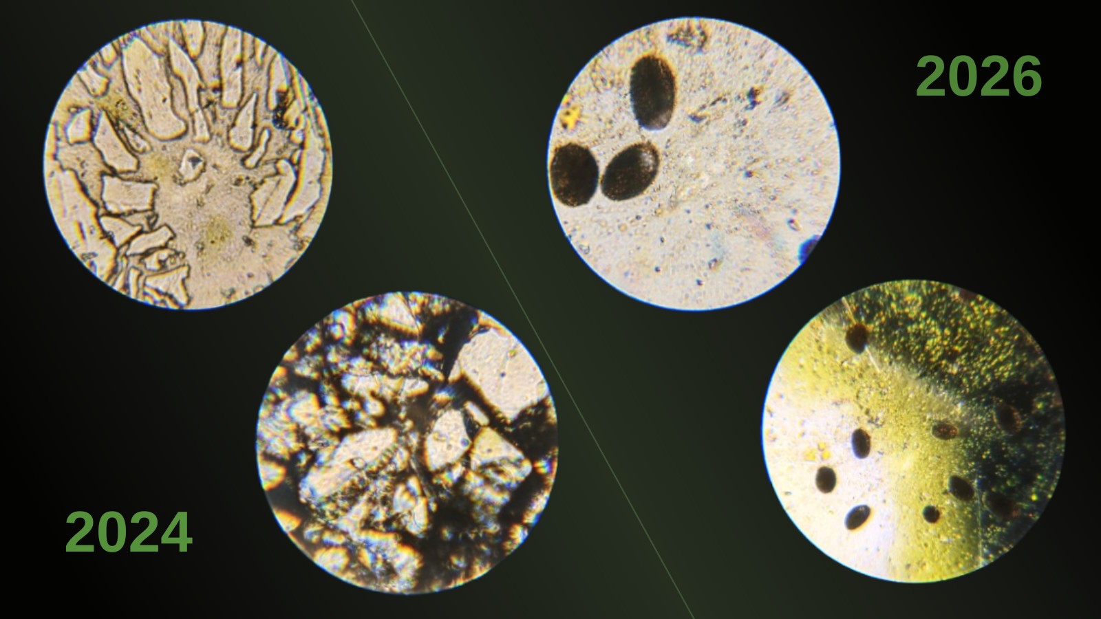
Previous attempt and comparison
I had performed a similar experiment approximately two years ago. However, due to an issue with the wet mount preparation, the cover slip detached, and I attempted to fix it using super glue. This likely altered the chemical composition of the sample, especially the sugar solution and pollen interaction, resulting in different visual outcomes compared to the present experiment.
Conclusion
Overall, these experiments provided a valuable opportunity to practice basic microscopy techniques and explore my interest in the microscopic world. Each attempt gives me a better understanding of what can be observed under the microscope and keeps my curiosity going.
References
->Microscopy guide used for educational purposes
->Personal observations and experiments
Stay Informed
Join our community of medical professionals and researchers. Get the latest insights delivered to your inbox.