Paleopathologies in non-avian dinosaurs
As today is Halloween, we needed at least one post to get us in the mood for spooky season. And, what is something we tend to associate with this time of year? Skeletons, of course. And what is known entirely from skeletons? Dinosaurs (specifically non-avian ones).
Although they are often seen as movie monsters, non-avian dinosaurs were not that different from many of the animals we have today, and, as such, endured many of the same kinds of hardships as modern animals and even humans do. So, on the night on which spirits walk among the living, let's bring back the ghosts of these long-dead animals in order to take a closer look at what they went through.
1. Tail amputation
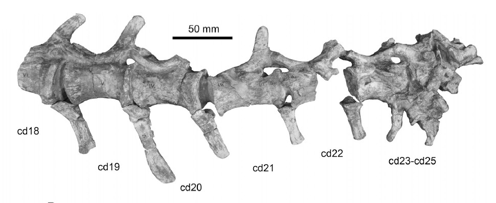
Discovered in 2008, in South Africa, and described in 2013 by Butler et al., specimen BP/1/6771 is most likely a dinosaur from the genus Massospondylus (whose name means “longer vertebra”), which lived in the Early Jurassic from around 201 to 184 million years ago. What is most notable about this animal is that its last three caudal vertebrae (numbers 23-25) are fused together, with signs of reactive bone (which is the growth of new bone as a result of either an injury or chronic irritation).
Figure from Butler et al. 2013, showing the traumatic ossification of the last 3 caudals.
What is more, although caudals 18-25 were found in perfect articulation, there were no signs of any other. Although no complete tail is known for Massospondylus, assuming it had a similar number of tail vertebrae to other basal Sauropodamorphs such as Plateosaurus and Lufengosaurus, then this animal lost about half of its tail. Although no definitive cause for the amputation is given in the paper, the team speculates it was most likely caused by a failed attack from a hungry theropod, such as Dracovenator, a contemporary of Massospondylus in the Elliot Formation of early Jurassic South Africa.
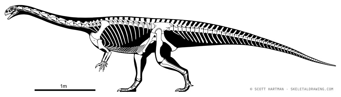
Skeletal reconstruction of Massospondylus by Scott Hartmann, showing the normal length of the tail.
2. Respiratory infection
Specimen MOR 7029, collected in 1990 in Montana, represents an as of yet indeterminate Diplodocine sauropod (a group which contains animals like Barosaurus, Supersaurus (whose main claim to fame is being a contender for the longest non-colonial organism ever, with lengths of 40 meters being suggested for some individuals) and the iconic Diplodocus) from the late Jurassic Morrison Formation (dated to around 155-145 million years ago) is one of the few dinosaur specimens known to have suffered from an infection to something else other than bone. By looking at the pathologies of the cervical vertebrae the team of scientists was able to determine that this individual, nicknamed “Dolly”, suffered from airsacculitis, a condition represented by the inflammation of the air sacs, which is also observed in modern day birds.
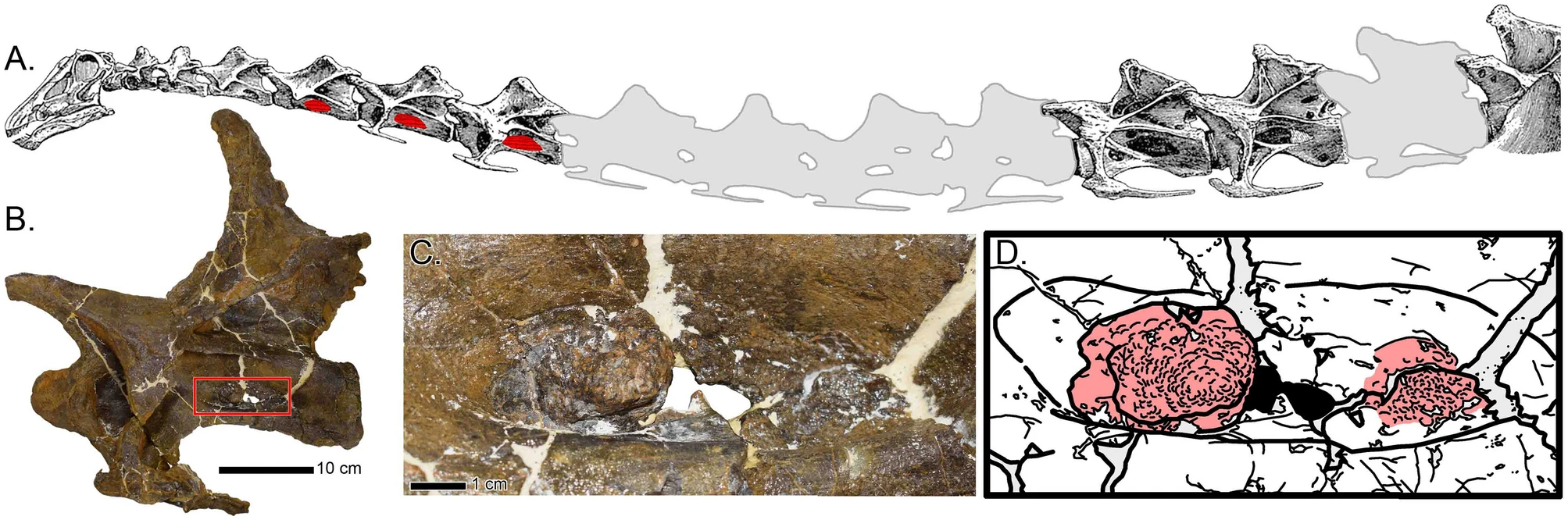
Firgure from Woodruff et al. 2022. A represents the neck of Diplodocus, with non-preserved elements in light gray and pathological structures in red, B is a picture of the specimen's fifth cervical, with the red box highlighting the pathological structures, shown in greater detail in C. D is an interpretative drawing of C.
Although the authors cannot say for certain what caused it, they have proposed several hypotheses, including pneumoconiosis (the inhalation of dust or other airborne particles), chlamydiosis (caused by Chlamyidia, a type of bacteria) or aspergillosis (caused by the fungal genus Aspergillus).
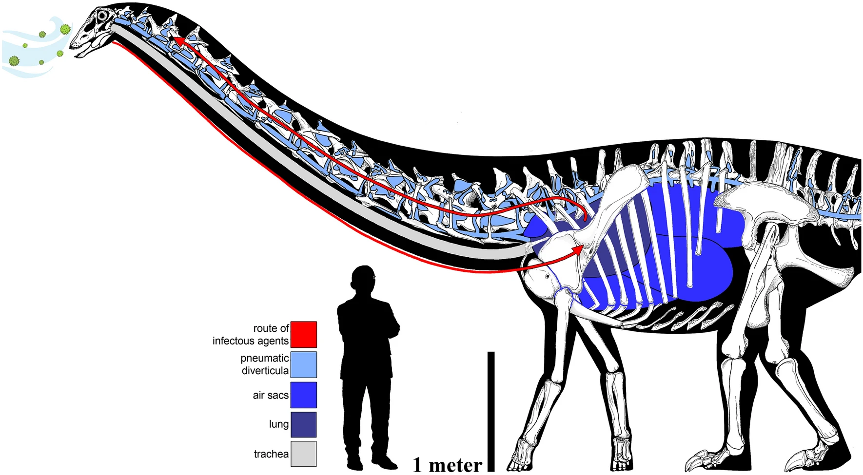
Another figure from Woodruff et al. 2022, showing a potential route of infectious the infectious pathway. The skeletal of the Diplodocine Galeamopus pabsti by Francisco Bruñén Alfaro is scaled to the proportions of MOR 7029.
3. The oldest dinosaur
ZPAL MgD-III/3, a specimen tentatively assigned to Gobihadros, a Hadrosauriform found in the Bayanshiree Formation of Mongolia during the early part of the Late Cretaceous, around 96 to 84 million years ago, is particularly interesting as it is one of the oldest known dinosaurs on record (not in terms of geologic age, but in terms of the age of the individual animal). The specimen was found with abnormal calcium deposits on two of its caudal vertebrae as well as on the first phalanx of the fourth digit of the left foot. These deposits are consistent with a disease called Calcium pyrophosphate deposition disease (CPPD for short), characterized by the accumulation of calcium pyrophosphate dihydrate crystals in the joint cartilage, thought to be caused by increased levels of ATP.
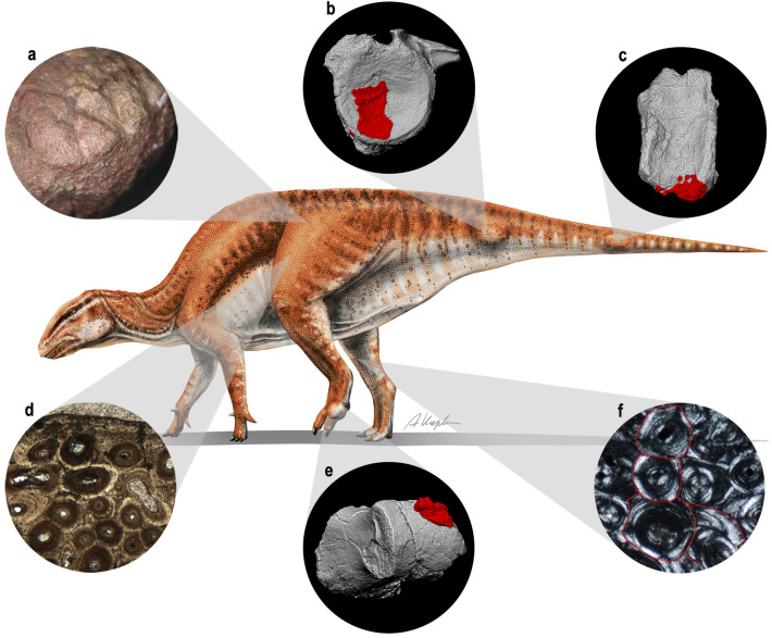
Figure from Słowiak et al. 2021 with reconstruction by A. Kapuścińska showing the various pathologies of the specimen.
Due to the fact CPPD is incredibly rare in human patients under 55 years old, the lines of arrested growth present in the cross-section of the femur as well as its very large size when compared to other specimens of Gobihadros, the researchers have thus come to the conclusion it was one of the oldest dinosaurs ever discovered, although no precise age is given.
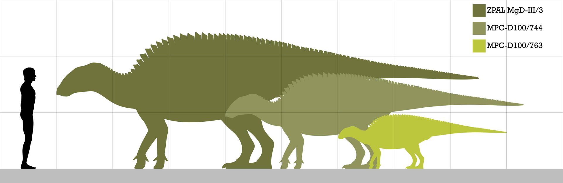
Size comparison between various specimens of Gobihadros by PaleoNeolitic, showing the large size of ZPAL MgD-III/3
4. Allosaurus, Allosaurus and more Allosaurus
Known as the “Lion of the Jurassic”, Allosaurus has gained the reputation of being reckless in the paleo-community due to the many, many injuries found on specimens of the genus. The first paleopathologies were noticed by Kristen Petersen et. al in 1972, which included two broken ribs, distorted joints, several fusions in some tail vertebrae an amputated chevron (bone on the underside of the tail) and toe bone, bony outgrowths on another toe bone and several injuries attributed to infections. There are three particular cases worth highlighting though.
The first is “Big Al” (specimen MOR 693), an individual of the species Allosaurus jimmadseni (I am specifying the species here because there are 3 other species of Allosaurus currently recognized as valid, those being A. fragilis, which is the type species, A. europaeus, and A. anax, the largest, formerly thought to be a separate genus Saurophaganax, although that’s a story for another time), found in the Morrison formation of Wyoming, which has 19 DIFFERENT PATHOLOGIES, described in detail by Hanna in 2002, including severe swelling of the first bone of the middle toe of the right foot, likely caused by an infection as well as an infection in the fifth metatarsal of the right foot would have caused even more swelling, probably causing the individual to limp as it walked. The first bone of the second finger was also infected, causing it to swell. And, to add insult to injury, this wasn’t even the last infection, as the fourth rib was also infected. Other pathologies were less serious, including growths on several vertebrae, as well as a healed chevron.
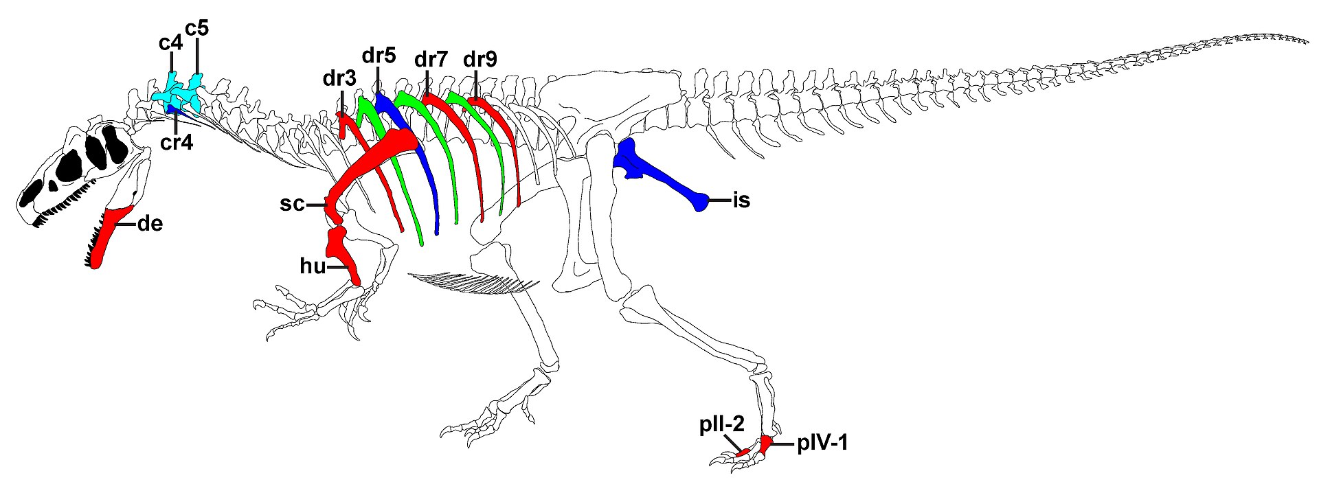
Pathologies of another individual belonging to Allosaurus jimmadseni named "Big Al II". Figure from Foth et al. 2015.
The other two injuries involve another one of the Morrison Formation’s most iconic dinosaurs: Stegosaurus, a dinosaur known for its spiked tail, and, thanks to two unfortunate Allosaurus specimens, we know it was put to good use. The first, described by Carpenter et al in 2005 is an Allosaurus caudal with a partially healed puncture wound, thought to have been caused by the thagomizer of a Stegosaur. However, it seems the predator didn’t live particularly long after its encounter, as there are not many signs of bone remodeling. However, if you thought that was bad, wait ‘till you see the “crotch shot” described by Bakker et al., which, as the name implies, is a fossil of an Allosaurus pubic boot that was pierced by the tail spike of a Stegosaurid. Although the animal appears to have survived the strike, it probably died of an infection soon after.
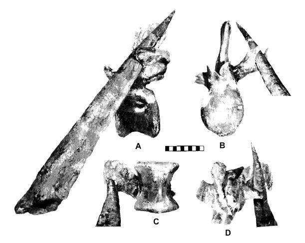
Figure from Carpenter et al. 2005 showing evidence that a Stegosaurus thagomizer caused the wound to the caudal.
5. Brutalized arms
Allosaurus wasn’t the only Jurassic theropod with multiple pathologies in a single individual. Dilophosaurus, a predator from the Early Jurassic (between around 195 and 184 million years ago) of Arizona who you probably know from its very inaccurate depiction in Jurassic Park (which differs from the real animal in several key ways, namely lacking the frill and ability to spit venom, but also being much larger, around 7m long and 400kgs in weight) had an individual that also endured large amounts of punishment.
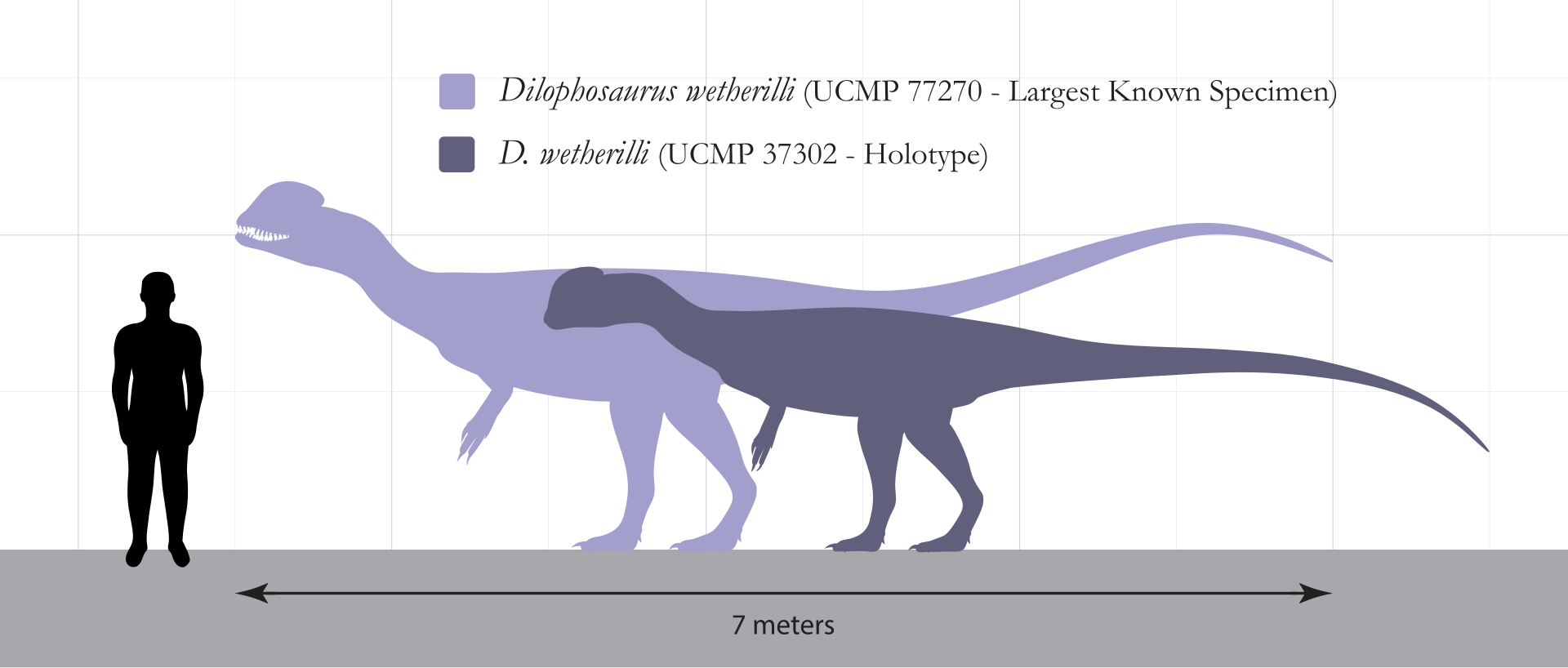
Size comparison between the two specimens of D. wetherilli and a human by SlvrHwk. The specimen discussed below is the holotype.
Although its injuries were first noticed by Welles, the same person who named the genus, the pathologies would be described in detail by Senter, Juengst and Heymann in 2016. They found that both of the holotype specimen’s arms were badly damaged, to a degree not seen in any other dinosaur. Its left arm had a fractured scapula and radius, as well as fibriscesses (a reptilian equivalent of abscesses) in the ulna and the outer phalanx of the thumb. Meanwhile the right side had torsion of its humeral shaft, three bony tumors on its radius, a truncated (shortened) articular surface of its third metacarpal, and deformities on the first phalanx bone of the third finger, making it permanently deformed and unable to flex. The scientists argue that the torsion in its right arm is due to osteodysplasia, which is known in modern birds. The animal is in so much pain that it refuses to use one of its limbs (in the case of the individual here, its left arm), leading to torsion developing in the other. However, since all these injuries had healed, it is likely the animal survived for months, if not years after.
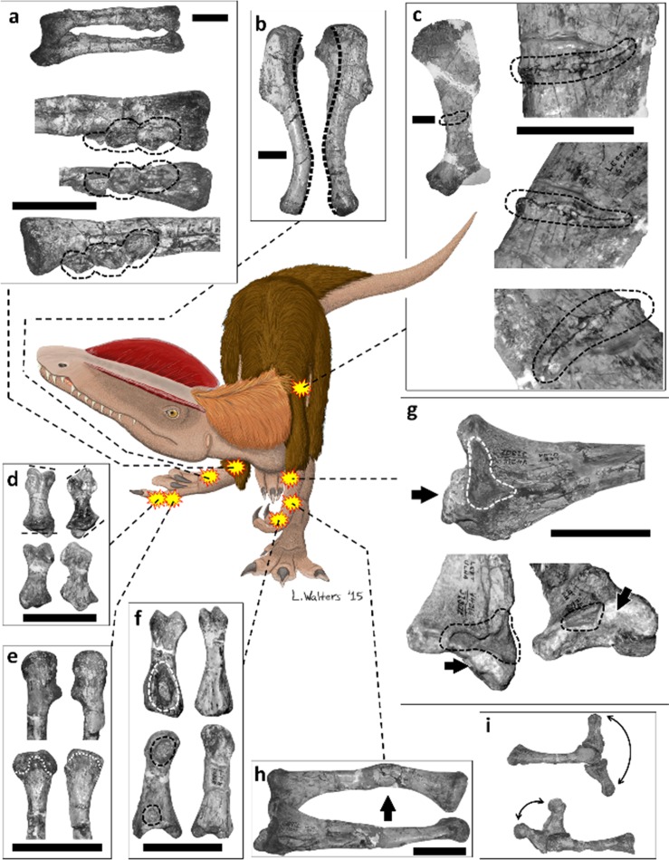
Figure from Senter, Juengst and Heymann 2016, showing the pathologies of the holotype of D. wetherilli.
6. Prehistoric cancer
Centrosaurus is one of the most well represented dinosaurs, known from the Dinosaur Park formation of Alberta, Canada, around 76 million years ago. It is so abundant that several bonebeds stretching for hundreds of meters containing thousands of specimens of all ages have been found, proving they lived together in large heards, which allowed individuals that wouldn’t survive out on their own make it.
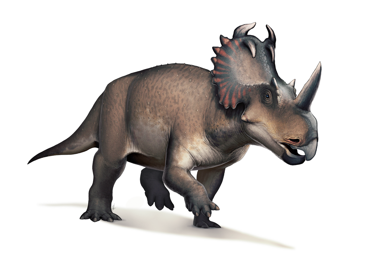
Life reconstruction of Centrosaurus apetrus by Fred Wierum.
Perhaps the clearest example is an individual whose right fibula had an unusual shape, which at first led paleontologists to suspect it was caused by a fracture that didn’t heal properly, however they later found that the dinosaur was suffering from osteosarcoma, a type of cancer which in humans primarily affects the long bones of teens and adults. The tumor was quite advanced, probably causing the dinosaur to limp. However, due to it living in a heard alongside other members of its kind, it survived for way longer than it would’ve alone, with this individual dying in a flood alongside the rest of its heard.
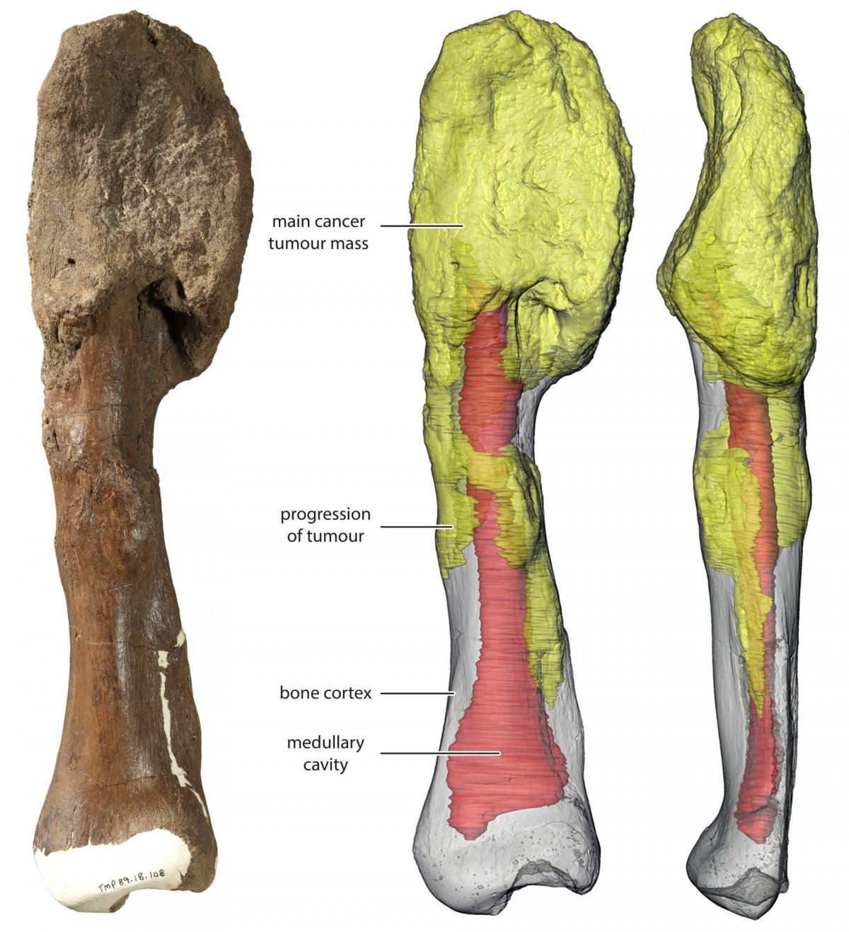
3D reconstruction of the Centrosaurus fibula. The main tumour mass is at the top of the bone in yellow; gray is the normal bone and red denotes the medullary cavity.
© Royal Ontario Museum/McMaster University
7. Failed hunt
There is no dinosaur that captures the public’s imagination quite like Tyrannosaurus rex, the “king of the dinosaurs”. However, what people forget sometimes is that most of today’s large predators have successful hunts less than 50% of the time, and T. rex was likely no different. In fact, we even have evidence for it. Hailing from the Hell Creek formation of south Dakota, deposited at the tail end of the Cretaceous around 66 million years ago is a hadrosaur caudal, most likely belonging to Edmontosaurus annectens (which sidenote, is known from several incredibly well preserved “mummies” allowing us to know things about its life appearance such as it possessing a fleshy crest on top of its head, or that it had hooves on both its front and back legs) with a large tooth stuck inside, belonging to none other than the Tyrant lizard king.
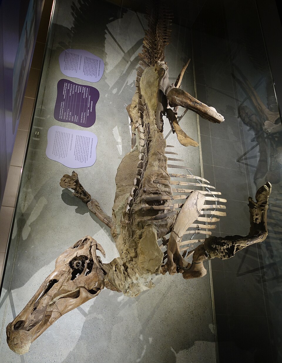
The Senckenberg mummy, on display in the Senckenberg museum in Frankfurt am Main. Photo by Jens Lallensack.
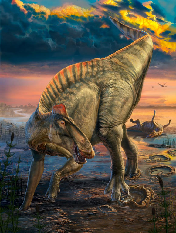
Life reconstruction of Edmontosaurus annectens by Dani Navarro, with insights from two new mummies described by Sereno et al. in 2025.
We are certain the hadrosaur managed to get away, however because there are signs of the bone healing around the tooth. This discovery was also the final nail in the coffin for the (absolutely bogus) theory that had been floating around in the 90s and early 2000s arguing T. rex was a scavenger. It really goes to show how important even a single discovery can be.
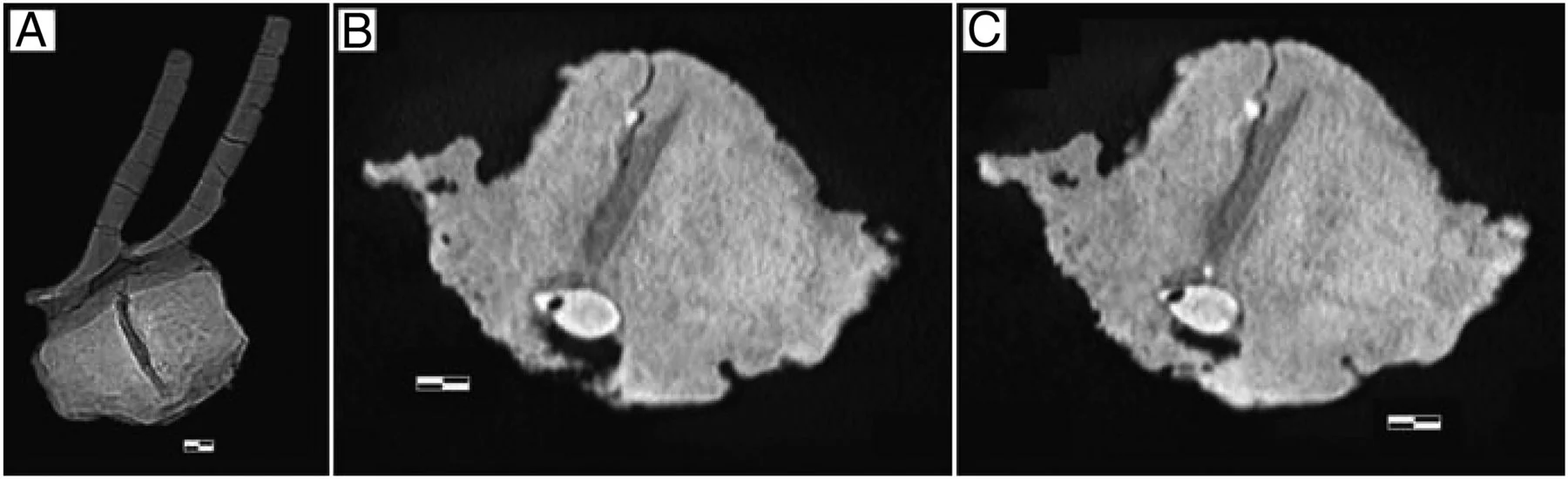
CT scans of the tooth embedded in the vertebra from DePalma et al. 2013.
8. The mother if all injuries
And, speaking of T. rex, I’ve saved the best (or worst depending on how you look at it) for last: NHMAD 2020.00001, also known as “Stan”. This guy truly had it all: a broken rib that became infected, a broken neck, leading to the fusion of two of his cervicals and if all that wasn’t enough, he even suffered a bite to the braincase from another T. rex.
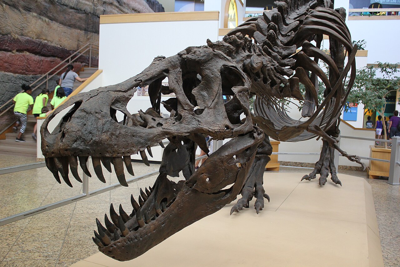
Cast of Stan at the New Mexico Museum of Natural History.
Although it’s likely his many injuries lead to his demise, the fact he was able to survive with all these injuries long enough for them to heal is a testament to just how resilient of an animal Tyrannosaurus was.
So, in the end, what can we learn from all these pathologies? For one, they can provide valuable details into the lives of dinosaurs, such as the heard behaviour of Centrosaurus, the predator-prey relationship between animals like Allosaurus and Stegosaurus or conspecific agression in Tyrannosaurus. It also shows us how several conditions seen in animals today, such as CPPD, osteodysplasia, osteosarcoma and airsacculitis are much more ancient than previously though. And, perhaps most importantly, it reminds us that non-avian dinosaurs were just animals, no different from the ones that exist today.
https://www.nature.com/articles/s41598-022-05761-3
https://pmc.ncbi.nlm.nih.gov/articles/PMC8196189/
https://www.researchgate.net/profile/Kenneth-Carpenter-2/publication/314890308_Evidence_for_Predator-_Prey_Relationships_Examples_for_Allosaurus_and_Stegosaurus/links/58c70893a
https://studio.m-anage.com/gsa/2014AM/webprogram/Paper247355.html6fdcc550c94d80a/Evidence-for-Predator-Prey-Relationships-Examples-for-Allosaurus-and-Stegosaurus.pdf?wpmobileexternal=true
https://peerj.com/articles/940/
https://pmc.ncbi.nlm.nih.gov/articles/PMC4765892/
https://pmc.ncbi.nlm.nih.gov/articles/PMC7983758/
https://www.pnas.org/doi/full/10.1073/pnas.1216534110
https://web.archive.org/web/20190404044028/https://bhigr.com/pages/info/info_stan.htm
Stay Informed
Join our community of medical professionals and researchers. Get the latest insights delivered to your inbox.